CT Coronary Angiography vs Traditional Coronary Angiography: Which Test Should Be Done First?- By Cardiologist, ONUS Robotic Hospitals
CT Coronary Angiography vs Traditional Coronary Angiography: Which Test Should Be Done First?- By Cardiologist, ONUS Robotic Hospitals
Chest pain, breathlessness, heaviness in the chest, sweating, or pain spreading to the left arm, jaw, neck, or back can be warning signs of heart disease. But many patients are confused: Should we go for CT Coronary Angiography first or Traditional Coronary Angiography?
The answer depends on the patient’s symptoms, risk level, emergency status, ECG findings, blood test results, and clinical condition.
For many stable chest pain patients, CT Coronary Angiography is used as a first-line test to assess coronary artery disease. Traditional invasive angiography is usually considered when CT findings are significant, tests are inconclusive, or treatment like angioplasty or stenting may be needed. NICE guidance recommends CT coronary angiography as first-line investigation when stable angina cannot be excluded by clinical assessment alone, while invasive coronary angiography is generally offered when CT findings are significant or further imaging is inconclusive.
In this video, Dr. Syeda Saba, Senior Clinical Cardiologist at ONUS Robotic Hospitals, explains the difference between CT Coronary Angiography and Traditional Coronary Angiography, when each test is useful, and when chest pain becomes a medical emergency.
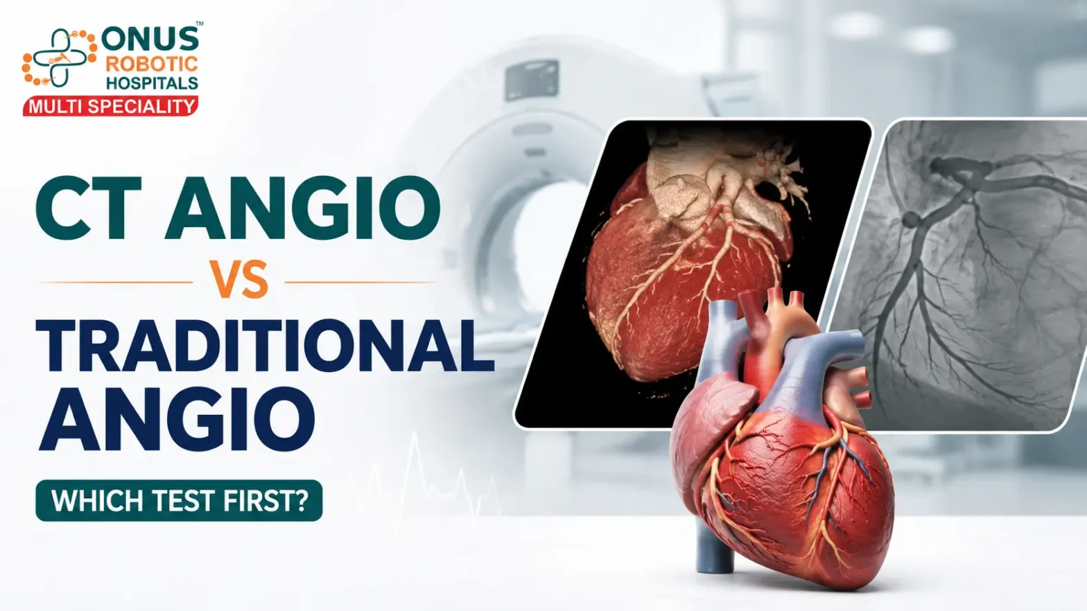
What Is CT Coronary Angiography?
CT Coronary Angiography, also called CT Coronary Angio or CCTA, is a non-invasive heart scan. It uses CT scan technology and contrast dye injected through a vein to create detailed images of the coronary arteries.
It helps doctors assess:
Coronary artery narrowing Plaque buildup Possible heart blockage Low to intermediate risk chest pain Stable chest pain evaluation Need for further cardiac testing
A CT coronary angiogram does not require tubes to be placed inside the body. Mayo Clinic explains that CCTA is a CT-based angiogram that does not need tubes placed inside the body and may help determine whether someone needs cardiac catheterization.
What Is Traditional Coronary Angiography?
Traditional Coronary Angiography is an invasive cath lab procedure. A thin tube called a catheter is inserted through a blood vessel, usually from the wrist or groin, and guided to the heart arteries. Contrast dye is injected to see the coronary arteries clearly.
This test is useful when:
There is strong suspicion of significant blockage CT coronary angiography shows major narrowing Stress test or other tests are abnormal Symptoms are high-risk Emergency heart attack care is needed Angioplasty or stenting may be required
Mayo Clinic notes that during a coronary angiogram, angioplasty and stenting can be done to open blocked arteries. A narrowing or blockage cannot be treated during a CT angiogram; if CT shows a blocked or narrowed artery, the catheter type of angiogram must be done.
CT Coronary Angio vs Traditional Angiogram: Main Difference
The main difference is that CT Coronary Angiography is mainly a diagnostic, non-invasive scan, while Traditional Coronary Angiography is an invasive cath lab procedure that can also allow treatment.
CT Coronary Angiography
Non-invasive scan Done using CT machine Contrast through vein Useful for stable chest pain evaluation Can show plaque and narrowing Cannot perform stenting Usually outpatient or short-duration test
Traditional Coronary Angiography
Invasive cath lab procedure Catheter inserted through wrist or groin Contrast injected directly into coronary arteries Useful when blockage is suspected or confirmed Can be followed by angioplasty or stenting Needed in emergency heart attack cases Requires cath lab setup and monitoring
Which Test Is Done First?
For many stable chest pain patients, CT Coronary Angiography may be considered first, especially when the patient is low to intermediate risk and not in emergency condition. The 2021 AHA/ACC Chest Pain Guideline provides an evidence-based approach for evaluating acute and stable chest pain, emphasizing risk stratification before selecting diagnostic tests.
However, if the patient has severe chest pain, ECG changes, high troponin levels, unstable symptoms, or suspected heart attack, emergency cardiac evaluation is needed. In such cases, doctors may proceed directly with urgent treatment pathways, including invasive angiography when required.
Stable Chest Pain vs Emergency Chest Pain
Stable Chest Pain
Stable chest pain may occur with exertion, walking, climbing stairs, stress, or physical activity and may improve with rest. These patients are usually evaluated based on risk factors, ECG, Echo, blood tests, and clinical judgment.
CT Coronary Angio may be useful in stable patients to assess coronary artery disease non-invasively.
Emergency Chest Pain
Emergency chest pain may be severe, sudden, prolonged, or associated with sweating, breathlessness, dizziness, fainting, nausea, or pain spreading to the arm, jaw, neck, or back.
Do not wait for routine testing if these symptoms are present. Seek emergency cardiac care immediately.
When CT Coronary Angiography Is Useful
CT Coronary Angio may be useful when:
Chest pain is stable Risk level is low to intermediate ECG and blood tests are not showing emergency heart attack Doctor wants to assess coronary arteries non-invasively Stress test is inconclusive There is a need to rule out significant coronary artery disease Patient needs anatomical assessment of heart arteries
A “CT-first” approach has been supported in stable chest pain pathways to help determine which patients may need revascularization.
When Traditional Coronary Angiography Is Needed
Traditional angiography may be needed when:
CT Coronary Angio shows significant narrowing Symptoms suggest high-risk heart disease ECG shows ischemic changes Troponin blood test is positive Patient has unstable angina Heart attack is suspected Angioplasty or stenting may be needed Other tests are inconclusive but symptoms are concerning
Traditional angiography is also important because treatment can be performed during the same procedure if required.
Role of ECG, 2D Echo, Blood Tests, CT Scan and Cath Lab
Heart disease evaluation is not based on one test alone. Doctors combine symptoms, risk factors, examination, and investigations.
CT coronary angio helps visualize heart arteries non-invasively and detect plaque or narrowing.
Traditional Coronary Angiography
Traditional angiography gives direct visualization of coronary arteries and allows angioplasty or stenting when needed.
When Chest Pain Becomes an Emergency
Chest pain becomes an emergency when it is associated with:
Severe chest heaviness Sweating Breathlessness Dizziness Fainting Nausea or vomiting Pain spreading to left arm Pain spreading to jaw Pain spreading to neck or back Chest discomfort lasting more than a few minutes Known diabetes, BP, cholesterol, smoking, or previous heart disease
If you suspect a heart attack, do not delay. Reach emergency care immediately.
Expert Cardiac Care at ONUS Robotic Hospitals
At ONUS Robotic Hospitals, patients with chest pain, breathlessness, suspected heart disease, stable chest pain, emergency chest pain, heart blockage, BP, diabetes-related heart risk, and cardiac symptoms receive timely evaluation and personalized care.